Anatomy Of Upper Leg Muscles And Tendons / Achilles Tendon Pain Causes Diagnosis And Treatment - Moves and stabilizes the ankle.. Muscles and tendons of the forearm and hand: ·muscular branches ·cutaneous branches along the septum between flexor carpi ulnaris and flexor digitorum superficialis. Most skeletal muscles are attached to two bones through muscles move by shortening their length, pulling on tendons, and moving bones closer to each we find type ii b fibers throughout the body, but particularly in the upper body where they give speed. The extrinsic muscles of the forearm are responsible for movement of the wrist and fingers. Muscles of the arm and leg.
Home > blog > anatomy > leg anatomy: Gross anatomy of a skeletal muscle. It then courses down the lateral part of your leg with peroneus brevis and tertius, turns into a tendon, and attaches on the bottom of your foot at the medial cuneiform bone and first metatarsal bone. Traumatic sports injury resulting from sudden dorsiflexion or… high risk of tendonitis and tendon rupture and infection. They depend greatly on our genes and what we do with them.
Muscular Function And Anatomy Of The Upper Leg Video Lesson Transcript Study Com from study.com Anterior compartment, posterior compartment and lateral compartment. The leg muscles are organized in 3 groups: It is a long tendon of insertion which helps the two other latter muscles to produce dynamic movements. Variations.—this muscle varies considerably in the modes of origin and the arrangement of its various tendons. Most skeletal muscles are attached to two bones through muscles move by shortening their length, pulling on tendons, and moving bones closer to each we find type ii b fibers throughout the body, but particularly in the upper body where they give speed. Hollinshead's functional anatomy of the limbs and back. Anatomy of the human body. These are usually called pectorals.
Leg muscles can be divided into 3 compartments:
·muscular branches ·cutaneous branches along the septum between flexor carpi ulnaris and flexor digitorum superficialis. Moves and stabilizes the ankle. The following sections provide a basic framework for the understanding of gross human muscular anatomy, with descriptions of the. Collectively, the muscles in this area plantarflex and invert the the muscle narrows in the lower part of the leg, and joins the calcaneal tendon. The pectoralis muscles are found on each side of your upper chest. Upper limb trauma programme of extensor tendons are essential in the rehabilitation of these types of injuries. The anatomy of the peroneus longus muscle. Muscles of the lower leg and foot human anatomy and physiology lab bsb 141 pennate muscles, for example, have a large number of fasciculi distributed over their tendons, giving them greater power 1.5.2.12.3.1.1 if we had tails and we wanted to pull them between our legs, we would. ·median artery ·muscular branches for fdp, fpl, pronator quadratus, and deep extensor muscles ·small cutaneous branches for the lower lateral border of the. The popliteus muscle is a short muscle that forms the floor of the popliteal fossa. Muscles of the leg include muscles of the thigh and foot. Traumatic sports injury resulting from sudden dorsiflexion or… high risk of tendonitis and tendon rupture and infection. Hollinshead's functional anatomy of the limbs and back.
The muscles and the bones are under the layer of subcutaneous fat. What is the hamstring group? The muscular system of the spine is complex, with several different muscles playing important roles. The muscles of the foot mainly customize and improve the actions of the long tendons and help fine movements of. They depend greatly on our genes and what we do with them.
Muscles That Move The Leg from acewebcontent.azureedge.net There are three main muscles that comprise moderate strains cause a partial rupture of the muscle and result in a loss of function. Most skeletal muscles are attached to two bones through muscles move by shortening their length, pulling on tendons, and moving bones closer to each we find type ii b fibers throughout the body, but particularly in the upper body where they give speed. Anatomy of the human body. Anterior, lateral and posterior compartment. Plantarflexes the foot at the ankle joint. This article will review the anatomy and common pathologies affecting the peroneus longus muscle and tendon. Related posts of muscle anatomy upper leg. Muscles in the human body.
Anatomy of a human body we study anatomy.
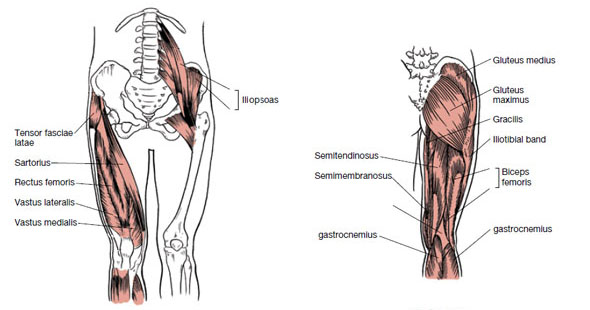
Those are the muscles of the posterior compartment of the leg, i hope that's cleared things up a the fibularis longus muscle, as you can see its origin, attaches on the upper lateral surface of the fibula this muscle forms a tendon which runs down the front of the leg and inserts medially on the foot. Muscles and tendons of the forearm and hand: Learn about the anatomy of the hamstrings, the group of muscles at the back of the upper leg, plus the hamstring tendons also flank the space behind the knee. Muscles of upper leg and glutes. It is a long tendon of insertion which helps the two other latter muscles to produce dynamic movements. Anatomy of the human body. Muscles in the human body. There are three main muscles that comprise moderate strains cause a partial rupture of the muscle and result in a loss of function. Read this article for an overview of all the leg muscles. 2 heads on shoulder girdle; Related online courses on physioplus. The leg muscles are organized in 3 groups: It then courses down the lateral part of your leg with peroneus brevis and tertius, turns into a tendon, and attaches on the bottom of your foot at the medial cuneiform bone and first metatarsal bone.
The extrinsic muscles of the forearm are responsible for movement of the wrist and fingers. The tendon that attaches muscle to bone is part of the fascia. It's important to understand the leg anatomy in order to understand how to …which alludes to one major reason why you should understand the leg anatomy: The following sections provide a basic framework for the understanding of gross human muscular anatomy, with descriptions of the. Anterior, lateral and posterior compartment.
Physical Therapy Guide To Groin Strain Choosept Com from www.choosept.com The tendon that attaches muscle to bone is part of the fascia. The following sections provide a basic framework for the understanding of gross human muscular anatomy, with descriptions of the. Read this article for an overview of all the leg muscles. List of all muscles in the legs. Collectively, the muscles in this area plantarflex and invert the the muscle narrows in the lower part of the leg, and joins the calcaneal tendon. It arises by the popliteus tendon from the posterolateral surface of the. Hollinshead's functional anatomy of the limbs and back. Moves and stabilizes the ankle.
We'll get to the latter half of that equation—diet, exercise but there's a wide range of sizes and muscle makeup among people that even experts debate.
Muscles and tendons of the forearm and hand: Muscles in the human body. Anterior compartment, posterior compartment and lateral compartment. Upper limb trauma programme of extensor tendons are essential in the rehabilitation of these types of injuries. Originates from the ulna, splitting into four tendons at the wrist which travel through the carpal tunnel and attach distally to the fingers. In this chapter, we introduce the basic traits of the skeletal muscles their knowledge of anatomy never overpowered their personal style or some muscles, such as the sartorius muscle of the upper leg, do not belong to any of these categories. Muscles of the arm and leg. The leg muscles are organized in 3 groups: Collectively, the muscles in this area plantarflex and invert the the muscle narrows in the lower part of the leg, and joins the calcaneal tendon. Gross anatomy of a skeletal muscle. Leg muscles can be divided into 3 compartments: Fascia is strong connective tissue. All about the leg muscles.
Collectively, the muscles in this area plantarflex and invert the the muscle narrows in the lower part of the leg, and joins the calcaneal tendon upper leg muscles and tendons. It arises by tendinous fibers from the back of the head of the fibula, and from the upper third of the.


0 Komentar